Rahatsızlığınıza Uygun Tedaviyi Bulun
2 adımda hızlıca bulun
Başlamak için rahatsızlığınızı seçin
Tedavi Bul
Tedavi Alanlarımız
Nörolojik, ortopedik, pediatrik ve kardiyopulmoner rehabilitasyondan robotik tedaviye uzanan kapsamlı hizmet yelpazemiz.
Fizik Tedavi
Fizik Tedavi Nedir? Fizik tedavi, kas iskelet sisteminde gerek doğuştan gerekse sonradan gelişen fiziksel ve fonksiyonel bozuklukların tanı ve tedavisi üzerinde yoğunlaşan bir tıp alanıdır. Uzman fizyoterapistler eşliğinde uygulanan bu tedavi; ağrıyı azaltmayı, hareketi artırmayı ve yaşam kalitesini yükseltmeyi hedefler. Fizik Tedavide Hangi Yöntemler Uygulanır? Soğuk ve sıcak tatbiki: Coldpack, hotpack, enfraruj, parafin banyosu Derin ısı terapisi: Ultrason, kısa dalga diatermi, mikrodalga diatermi, ultraviyole Elektroterapi: Galvani, faradi, TENS, diadinami, interferans, lazer terapi İyontoforez: Ultrason veya elektroterapi ile ilaç geçişi Hidroterapi: Kaplıca tedavisi, havuz tedavisi, balneoterapi, fango uygulamaları Mekanoterapi: Mobilizasyon, manipülasyon, traksiyon, masaj, splint, korse, bandaj Tedavi edici egzersiz uygulamaları Ağrı kesici bantlama uygulamaları Fizik Tedavinin Amaçları Nelerdir? Ağrının azaltılması Kasların gevşetilmesi Dolaşımın olumlu yönde etkilenmesi İnflamasyonun giderilmesi Fonksiyonların düzenlenmesi ve hareketlerin artırılması Kasların koordinasyonunun sağlanması ve güçlendirilmesi İlaç ihtiyacının azaltılması Duruş bozukluklarının önlenmesi ve düzeltilmesi Fizik Tedavi Hangi Hastalıklarda Uygulanabilir? Akut ve kronik ağrı tedavisi Romatizmal hastalıkların takip ve tedavisi Ortopedik rehabilitasyon ve spor yaralanmaları Nörolojik ve nöromüsküler hastalıkların rehabilitasyonu Çocuk rehabilitasyonu Metabolik kemik hastalıkları (osteoporoz vb.) Doğumsal veya edinsel eklem ve kemik bozuklukları Kardiyak rehabilitasyon Geriatrik rehabilitasyon Pulmoner rehabilitasyon Fizik Tedavi Uygulamalarının Faydaları Nelerdir? Ağrı, uyuşukluk, karıncalanma ve güçsüzlüğün azalması Eklem hareket açıklığının tekrar kazanılması Gereksiz tıbbi ve cerrahi tedavilerin engellenmesi Daha az ilaç kullanımının sağlanması Hastalıkların kronikleşerek ilerlemesinin engellenmesi Yaşam kalitesinin artması Fizik Tedavi Kimlere Uygulanır? Fizik tedavi; çocuklardan yaşlılara, sporculardan masa başı çalışanlara kadar her yaş grubundaki bireyler için uygundur. Ağrısı, hareket kısıtlılığı veya fonksiyon kaybı olan herkes fizik tedaviden yararlanabilir. Fizik Tedavi Ne Kadar Sürer? Bir fizik tedavi seansı uygulanan yöntemlere göre genellikle 30 ile 60 dakika arasında sürer. Tedavi süresi hastalığın türüne ve şiddetine göre değişmekle birlikte hafif sorunlarda 2–4 hafta, kronik durumlarda 6–12 hafta sürebilir. Fizik Tedavi Sürecinde Dikkat Edilmesi Gerekenler Tedavi programına düzenli olarak katılın Evde uygulama programına sadık kalın Şikayetlerinizi fizyoterapistinizle paylaşın Ağrınızın artması durumunda derhal bildirimde bulunun İlaç değişikliklerini yalnızca hekim onayıyla yapın
Sporcu Rehabilitasyonu
Sporcu Rehabilitasyonu Nedir? Sporcu rehabilitasyonu, spor esnasında ya da antrenman sürecinde meydana gelen yaralanmalar sonrası bireylerin mümkün olan en kısa sürede yaralanma öncesi performans düzeylerine kavuşmasını sağlamak amacıyla uygulanan bilimsel tedavi sürecinin bütünüdür. Bu süreç yalnızca profesyonel sporcuları değil; sağlıklı kalmak amacıyla düzenli spor yapan bireyleri, sahne sanatçılarını ve bedenini mesleği için kullanan çeşitli meslek gruplarını da kapsar. Sporcu Rehabilitasyonunda Neler Amaçlanır? Tedavinin temel hedefleri şunlardır: Ağrının giderilmesi ve iltihabın kontrol altına alınması Kas kuvveti ve dayanıklılığın yeniden kazanılması Eklem hareket açıklığının normale döndürülmesi Propriyosepsiyon ve koordinasyonun yeniden eğitilmesi Spora özgü fonksiyonel becerilerin yeniden kazanılması Yeniden yaralanmaların önlenmesi Sporcu Rehabilitasyonu Özellikleri Nelerdir? Sporcu rehabilitasyonu, standart rehabilitasyondan farklı olarak daha agresif ve spora özgü bir program gerektirir. Her sporcunun yaralanması, branşı ve fiziksel özellikleri göz önünde bulundurularak bireyselleştirilmiş bir program hazırlanır. Tedavi ekibinde fizik tedavi ve rehabilitasyon uzmanı, fizyoterapist ve gerektiğinde branş uzmanı yer alır. Tedavi Yöntemleri Manuel terapi teknikleri Eklem mobilizasyonu ve stabilizasyonu Propriyoseptif ve nöromüsküler eğitim Kuvvetlendirme ve dayanıklılık egzersizleri Hidro terapi ve aqua egzersizler Elektroterapi uygulamaları (TENS, ultrason, lazer) Kinesiotape uygulaması Kimler Yararlanabilir? Sporcu rehabilitasyonundan; spor yaralanması geçiren profesyonel ve amatör sporcular, ameliyat sonrası spora dönmek isteyen bireyler ve beden performansı mesleği olan kişiler (dansçı, tiyatro sanatçısı vb.) yararlanabilir.
Kognitif (Bilişsel) Terapi
Kognitif (Bilişsel) Terapi Nedir? Kognitif (bilişsel) terapi, beyin hasarı, inme, travmatik beyin yaralanması veya nörolojik hastalıklar nedeniyle bozulan bellek, dikkat, algı, problem çözme ve yürütücü işlevler gibi bilişsel becerilerin yeniden kazanılmasını hedefleyen uzmanlaşmış bir rehabilitasyon yaklaşımıdır. ROMMER International'da ergoterapi temelli bilişsel değerlendirme ve müdahale programları uygulanmaktadır. Hangi Durumlar İçin Uygulanır? İnme (serebrovasküler olay) sonrası bilişsel bozukluklar Travmatik beyin yaralanması Nörolojik hastalıklar (MS, Parkinson, beyin tümörü vb.) Dikkat eksikliği ve konsantrasyon sorunları Bellek bozuklukları Yürütücü işlev bozuklukları (planlama, sıralama, karar verme) Görsel-uzamsal algı bozuklukları Değerlendirme Süreci Tedaviye başlamadan önce standardize bilişsel testler kullanılarak hastanın dikkat, bellek, yürütücü işlevler ve işlem hızı gibi alanlardaki güçlü ve zayıf yönleri belirlenir. Bu değerlendirme, kişiye özel terapi programının temelini oluşturur. Terapi Yöntemleri Kompenzatuar stratejiler (dış yardımcılar, liste yapma, alarm sistemleri) Yeniden öğrenme teknikleri (tekrar, aşamalı zorluklarda pratik) Bilgisayar destekli bilişsel eğitim programları Günlük yaşam aktivitelerinde bilişsel eğitim Çevre düzenlemesi ve uyarlanmış görevler Tedavinin Faydaları Düzenli bilişsel terapi ile hastalar; dikkat ve konsantrasyonlarını güçlendirebilir, kısa ve uzun dönem belleklerini geliştirebilebilir, günlük yaşamda bağımsızlıklarını artırabilir ve iş yaşamına ya da eğitime geri dönüşlerini kolaylaştırabilirler.
Yutma Bozukluğu Rehabilitasyonu
Yutma Bozukluğu (Disfaji) Rehabilitasyonu Nedir? Yutma bozukluğu (disfaji), besin ve sıvıların ağızdan mideye güvenli şekilde ulaşmasını engelleyen bir durumdur. Nörolojik hastalıklar, beyin hasarı, baş boyun kanserleri ve yaşlılığa bağlı gelişebilir. Yutma rehabilitasyonu; oral-motor kasların güç, koordinasyon ve hareketliliğini geliştirerek güvenli yutmayı yeniden kazandırmayı amaçlar. Kimler Etkilenir? İnme (felç) geçirenler Parkinson ve MS hastaları Beyin hasarı yaşayanlar Baş ve boyun kanseri tedavisi görenler Demans hastaları Yutma güçlüğü olan yaşlı bireyler Değerlendirme Tedaviye başlamadan önce konuşma-dil terapisti ve fizik tedavi uzmanı tarafından kapsamlı bir yutma değerlendirmesi yapılır. Gerektiğinde videofloroskopik veya endoskopik yutma değerlendirmesi uygulanabilir. Tedavi Yöntemleri Oral-motor egzersizler (çene, dil, dudak güçlendirme) Kompansatuar yutma stratejileri (çene tuck, baş rotasyonu) Besin kıvam ve tekstür modifikasyonu Termal-taktil stimülasyon Nöromüsküler elektriksel stimülasyon (VitalStim) Hasta ve aile eğitimi (aspirasyon önlemi) Hedefler Güvenli oral beslenmeye dönüş, aspirasyon pnömonisi riskinin azaltılması, beslenme tüpünden bağımsızlık ve yaşam kalitesinin iyileştirilmesidir.
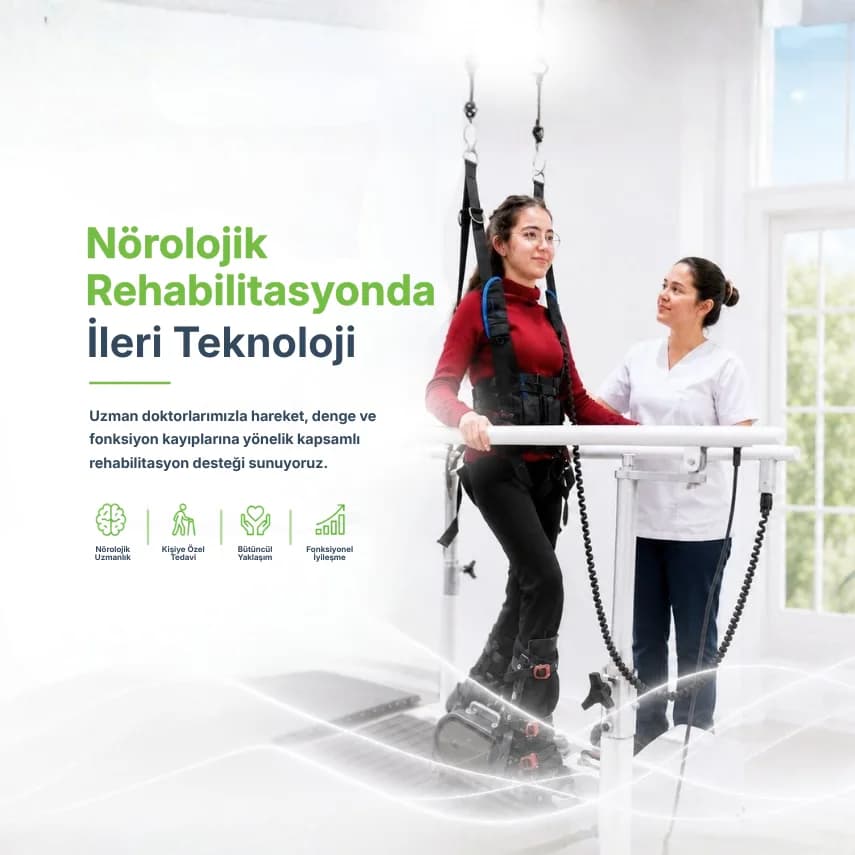
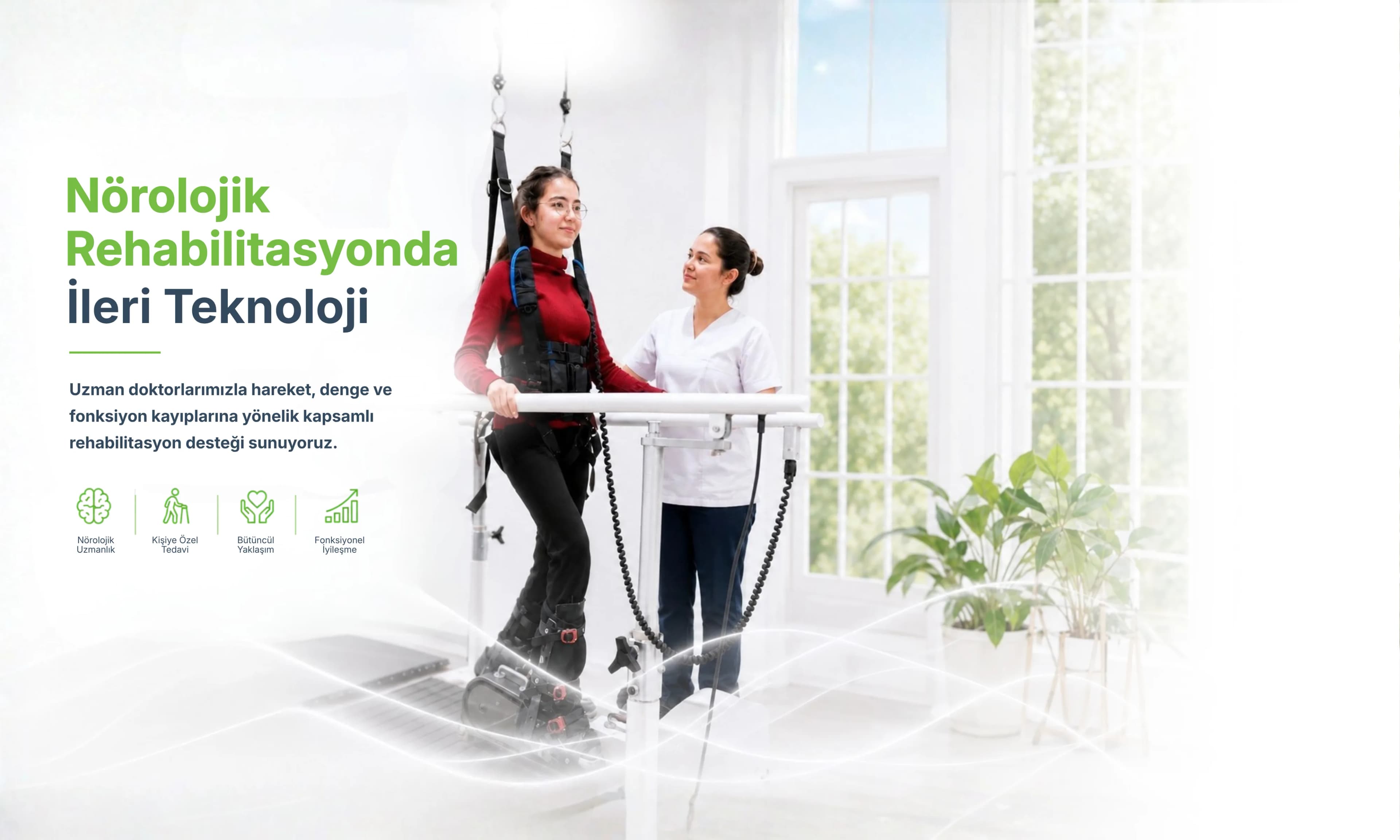
Yürüme ve Denge Bozukluğu Rehabilitasyonu
Yürüme ve Denge Bozukluğu Rehabilitasyonu Nedir? Yürüme ve denge, merkezi sinir sistemi, periferik sinir sistemi, kas-iskelet sistemi ve duyusal sistemlerin (görme, vestibüler, propriyosepsiyon) uyumlu çalışmasına dayanır. Bu sistemlerin herhangi birinde oluşan bozukluk; dengesizlik, düşme riski, güvensiz yürüyüş ve bağımsızlık kaybına yol açabilir. ROMMER International'da yürüyüş analizi ve gelişmiş denge eğitimi sistemleri ile kapsamlı değerlendirme ve tedavi sunulmaktadır. Neden Oluşur? İnme ve nörolojik hastalıklar Vestibüler bozukluklar (iç kulak problemleri) Parkinson hastalığı Multiple skleroz Periferik nöropati Ortopedik sorunlar (kalça, diz, ayak bileği) İlaç yan etkileri Yaşlılığa bağlı denge kaybı Değerlendirme Yürüyüş analizi, denge testleri (Berg Denge Skalası, TUG testi) ve kapsamlı nörolojik değerlendirme ile bireyin mevcut durumu ve düşme riski belirlenir. Tedavi Yöntemleri Denge eğitim platformları ve denge tahtası çalışmaları Yürüyüş yeniden eğitimi (treadmill, paralel bar, zemin eğitimi) Güçlendirme egzersizleri (alt ekstremite, core) Propriyosepsiyon ve duyu eğitimi Vestibüler rehabilitasyon Robotik yürüyüş eğitimi Düşme önleme stratejileri ve ev güvenliği düzenlemeleri Hedefler Güvenli ve bağımsız yürüyüşün yeniden kazanılması, düşme riskinin azaltılması ve yaşlı bireylerde topluma katılımın artırılması tedavinin temel hedefleridir.
Amputasyon Rehabilitasyonu
Amputasyon Rehabilitasyonu Nedir? Amputasyon rehabilitasyonu, üst veya alt ekstremite kaybı yaşayan bireylerin mümkün olan en yüksek işlevsellik ve yaşam kalitesine kavuşması için uygulanan kapsamlı rehabilitasyon programıdır. ROMMER International'da fizik tedavi uzmanı, fizyoterapist ve ergoterapi uzmanından oluşan multidisipliner ekip, her hastaya özel bir rehabilitasyon yol haritası belirler. Amputasyon Sonrası Rehabilitasyon Aşamaları Ameliyat öncesi hazırlık: Kas kuvvetlendirme, kondisyon geliştirme ve hasta eğitimi Akut dönem: Güdük bakımı, ödem kontrolü, ağrı yönetimi ve pansuman eğitimi Protez öncesi eğitim: Denge ve yürüyüş eğitimi, transfer teknikleri Protez uyum eğitimi: Protez kullanımı, bakımı ve günlük yaşama entegrasyonu Fonksiyonel rehabilitasyon: Merdiven çıkma, arazi yürüyüşü, mesleğe dönüş Rehabilitasyon Yöntemleri Güdük egzersizleri ve güçlendirme Denge ve propriyosepsiyon eğitimi Fantom ağrısı yönetimi (ayna terapisi, TENS) Protez ile yürüyüş analizi ve eğitimi Üst ekstremite amputasyonlarında el beceri eğitimi Günlük yaşam aktiviteleri eğitimi (ergoterapi) Ev modifikasyonu ve yardımcı cihaz önerileri Psikolojik Destek Uzuv kaybı bireyin hem fiziksel hem de psikolojik iyilik halini etkiler. Rehabilitasyon programımız, hastanın yeni beden imgesine uyum sürecini ve özgüven gelişimini destekleyecek psikolojik destek bileşenlerini de içermektedir.
Kas Hastalığı Terapisi
Kas Hastalıkları Nedir? Kas hastalıkları vücudun hareket etmesine yardımcı olan kasların kendine ait rahatsızlıklarıdır. Kas dokularının aralarında olan kas hücrelerinin yapı taşlarını veya işleyişini bozmakta ve faaliyetlerini de büyük oranda etkilemektedir. Bebeklikten çocukluk evresine, ergenlik, erişkinlik ve yaşlılığa kadar hayatın her evresinde her yaşta bireyde görülebilen yaygın bir hastalık türüdür. Kas Hastalıklarının Belirtileri Nelerdir? Oldukça yaygın görülen ve bireyi normal aktivitelerini yapmaktan alıkoyan kas hastalıklarının belirtileri şunlardır: Kalça ve çevresinde bulunan kaslar ortaya çıkacağından yürüme bozuklukları Kalkma ve yürümede zorluk çekme Küçük yaşlarda kas hastalığı ortaya çıkan çocuklar yürürken veya merdiven çıkarken sürekli kucağa alınmak ister Ergenlik döneminde yaşıtlarından geri kalma, yürümede değişme ve hareketlerinde farklılıklar görülür Omur ve çevresindeki kaslar tutulma yapacağından kolları kaldırmada, uzatmada ve hareket ettirmede güçlük yaşanır El ve ayaklar etkilenirse, yürürken takılma ve düşme (buna bağlı ayakkabıların çabuk eskimesi), el ile ince işleri yapamayacak durum gözlenir Nadir de olsa göz kapakları giderek aşağıya düşer, göz harekeleri kısıtlanabilir Yutma veya solunum kaslarında tutulma yaşanır Sık düşme, çabuk yorulma Kasları tutan hastalıklar Kas Hastalıkları Kimlerde Görülür? Kaslardaki anormallik sonucu, hafif belirtiler veren tipleri yanı sıra, şiddeti yüksek bir özre yol açan ve yaşam süresini azaltabilen çeşitleri de vardır: Musküler Distrofiler (Duchenne, Becker, FasioSkapuloHumeral, Limb-Girdle, Distal, Emery-Dreifuss) Konjenital Musküler Distrofiler Miyopatiler Miyotoniler Dermatomiyozit-Polimiyozit Kas Hastalıklarında Ergoterapinin Amaçları Nelerdir? Kas hastalıklarında rehabilitasyon yaklaşımlarının amacı, çocuğun ve ailesinin yaşam kalitesini yükselterek aktivitelere bağımsız bir şekilde katılımını sağlamaktır. Buna yönelik olarak yapılan uygulamaların amacı şöyledir: Kas kuvvetinin korunmasını sağlamak veya kuvvet kaybını geciktirmek Hastalığın farklı dönemlerinde eklemi desteklemek, korumak, fonksiyonu artırmak amacıyla uygun araç, gereç ve cihazlardan yararlanmak Yorgunlukla baş etme yöntemlerini öğretmek Eklem ve enerji koruma yöntemlerini öğretmek Fonksiyonel kapasiteyi artırarak günlük yaşam aktivitelerindeki bağımsızlığı sağlamak Ev rehabilitasyonu ile bireyin daha emniyetli ve daha az enerji sarf edeceği yaşam alanı sağlanır Koruyucu rehabi̇li̇tasyon yaklaşımlarıyla varolan fonksi̇yonel kapasi̇te daha uzun süre korunmaya çalışılır. Kas Hastalıklarında Ergoterapi Nasıl Uygulanır? Kas hastalıklarında ergoterapi, bireyin günlük yaşamını mümkün olduğunca bağımsız sürdürebilmesini hedefleyen bütüncül bir rehabilitasyon yaklaşımıdır. Süreç, hastanın kas gücü, hareket kabiliyeti, koordinasyonu ve fonksiyonel kapasitesinin detaylı şekilde değerlendirilmesiyle başlar. Ergoterapist, hastanın hangi hareketlerde zorlandığını, hangi aktivitelerde destek ihtiyacı olduğunu analiz ederek kişiye özel bir program oluşturur. Bu program yalnızca egzersizlerden ibaret değildir; aynı zamanda günlük yaşam aktivitelerinin nasıl daha verimli ve güvenli yapılabileceğini öğretmeye yönelik stratejiler de içerir. Örneğin, giyinme, yemek yeme veya kişisel bakım gibi temel aktivitelerde daha az enerji harcayarak daha fazla iş yapabilmeyi sağlayan teknikler öğretilir. Uygulama sürecinde, kas gücünü destekleyen fonksiyonel egzersizler, ince motor becerileri geliştiren çalışmalar ve çevresel düzenlemeler birlikte ele alınır. Ergoterapi yalnızca fiziksel kapasiteyi artırmayı değil, aynı zamanda bireyin yaşam kalitesini yükseltmeyi amaçlar. Bu nedenle terapi sürecinde yardımcı cihazlar (ortezler, özel tutacaklar, adaptif ekipmanlar) kullanılabilir. Ayrıca hastanın yaşadığı ortam da değerlendirilerek, ev veya iş yerinde yapılabilecek küçük düzenlemelerle hareket kabiliyeti artırılır. Bu yaklaşım sayesinde hasta, mevcut fiziksel sınırlarına rağmen günlük yaşamını daha bağımsız ve konforlu bir şekilde sürdürebilir. Kas Hastalarında Günlük Yaşam Aktiviteleri Nasıl Kolaylaştırılır? Kas hastalıklarında günlük yaşam aktivitelerinin kolaylaştırılması, hem fiziksel yükü azaltmak hem de bireyin bağımsızlığını korumak açısından büyük önem taşır. Bu süreçte en önemli adım, aktivitelerin hastanın mevcut kapasitesine göre yeniden düzenlenmesidir. Örneğin, uzun süre ayakta kalmayı gerektiren işler oturarak yapılacak şekilde adapte edilebilir. Enerji koruma teknikleri bu noktada devreye girer; yani kişi, gün içinde yapacağı işleri planlayarak gereksiz yorgunluğu önler ve daha verimli bir rutin oluşturur. Ayrıca hareketleri daha küçük parçalara bölmek, zorlayıcı aktiviteleri dinlenme aralarıyla desteklemek de günlük yaşamı ciddi ölçüde kolaylaştırır. Bunun yanı sıra yardımcı ekipmanlar ve çevresel düzenlemeler de büyük fark yaratır. Kavrama gücü azalan hastalar için özel tasarlanmış mutfak gereçleri, kaymaz yüzeyler veya kolay açılabilir kapaklar günlük işleri daha pratik hale getirir. Banyoda tutunma barları, yükseltilmiş oturma alanları veya kaydırmaz zeminler güvenliği artırır. Giyinme sırasında fermuar yerine cırt cırtlı veya elastik kıyafetler tercih edilmesi gibi küçük değişiklikler bile büyük kolaylık sağlar. Tüm bu düzenlemeler, kas hastalarının hem fiziksel olarak daha az zorlanmasını hem de psikolojik olarak daha bağımsız hissetmesini destekler.
Snoezelen Terapisi
Snoezelen Terapisi Nedir? Snoezelen terapisi, özel tasarlanmış çok duyusal bir odada ışık efektleri, rahatlatıcı sesler, farklı dokular, aromaterapi ve titreşimli yüzeyler aracılığıyla duyusal uyarım sağlayan terapötik bir yaklaşımdır. 1970'lerde Hollanda'da geliştirilen bu yöntem, nörolojik ve gelişimsel bozuklukları olan bireyler için etkili bir terapi ortamı sunar. Kimler İçin Uygundur? Otizm spektrum bozukluğu olan çocuklar ve yetişkinler Serebral palsi hastaları Down sendromlu bireyler Gelişimsel gecikmesi olan çocuklar Demans ve Alzheimer hastaları Kronik ağrı ve stres yaşayan bireyler Travma sonrası stres bozukluğu (TSSB) olan kişiler Duyusal işleme bozukluğu olan bireyler Snoezelen Odasında Ne Yer Alır? Renk değiştiren fiber optik ışıklar ve lamba topları Rahatlatıcı müzik ve doğa sesleri Titreşimli yatak ve yastıklar (vibroakustik) Farklı doku ve materyaller (dokunsal panel) Aromaterapi difüzörleri Projeksiyon görselleri ve balonlar Terapinin Faydaları Anksiyete ve ajitasyonun azaltılması Duyusal duyarlılığın düzenlenmesi İletişim ve sosyal etkileşimin desteklenmesi Ağrı algısının azaltılması Uyku kalitesinin iyileştirilmesi Genel iyilik hali ve yaşam kalitesinin artırılması
Sosyal Beceri Eğitimi
Sosyal Beceri Eğitimi Nedir? Sosyal beceri eğitimi, kişilerin başkalarıyla etkili iletişim kurma, duygu ve düşüncelerini uygun biçimde ifade etme, sosyal normlara uyum sağlama ve sağlıklı ilişkiler kurma becerilerini geliştirmeye yönelik yapılandırılmış bir terapi yaklaşımıdır. ROMMER International'da ergoterapi uzmanları tarafından uygulanan bu program; otizm, serebral palsi, Down sendromu, gelişim geriliği ve psikiyatrik bozuklukları olan bireyleri kapsamaktadır. Sosyal Yeterlilik Nedir? Sosyal yeterlilik üç boyutlu bir yapıda incelenir: sosyal uyum, sosyal performans ve sosyal beceri. Bu beceriler; özgüven, göz teması, konuşma sırası, empati ve uygun sosyal vurgulama gibi parametrelere bağlıdır. Kimler Yararlanır? Otizm spektrum bozukluğu olan çocuklar ve yetişkinler Down sendromlu bireyler Serebral palsili bireyler Gelişim geriliği olan çocuklar Sosyal kaygı ve fobisi olan bireyler Psikiyatrik bozukluğu olan bireyler (şizofreni, bipolar) Akran ilişkilerinde güçlük yaşayan çocuklar Program İçeriği Göz teması ve dikkat becerileri Sıra bekleme ve paylaşma Duyguları tanıma ve ifade etme Grupla oyun ve işbirliği Problem çözme ve çatışma yönetimi Öfke kontrolü teknikleri Günlük yaşam becerilerine entegrasyon Terapi Süreci Bireyin mevcut sosyal performansının detaylı değerlendirmesiyle başlanır. Grup çalışmaları, rol yapma uygulamaları ve gerçek yaşam senaryoları ile öğrenilen davranışların kalıcı hale gelmesi sağlanır. Aile de aktif olarak sürece dahil edilir.
Skolyoz Rehabilitasyonu
Skolyoz Rehabilitasyonu Nedir? Skolyoz, omurganın normalde olması gerektiği gibi düz olmaması; sağa veya sola yana doğru eğilmesi durumudur. Görülme sıklığı yaklaşık %2-4 olup kız çocuklarında erkek çocuklarına oranla çok daha sık görülmektedir. ROMMER International'da bilimsel temelli Schroth yöntemi ve çağdaş fizyoterapi yaklaşımları birleştirilerek kapsamlı bir skolyoz rehabilitasyon programı sunulmaktadır. Skolyoz Türleri İdiopatik skolyoz: En sık görülen tür; nedeni bilinmez, ergenlik döneminde sıkça ortaya çıkar Konjenital skolyoz: Doğuştan omurga gelişim bozukluğu Nöromüsküler skolyoz: Serebral palsi, kas distrofisi gibi nörolojik hastalıklara bağlı Dejeneratif skolyoz: İleri yaşta omurga dejenerasyonuna bağlı Belirtiler Omuzların ya da kalçaların eşit yükseklikte olmaması Öne eğilince sırtta belirgin asimetri Sırt ve bel ağrısı Bir kürek kemiğinin daha çıkıntılı görünmesi Konservatif Tedavi Yöntemleri Schroth metodu (özel solunum ve düzeltici egzersizler) SEAS (Scientific Exercise Approach to Scoliosis) egzersizleri Aktif stabilizasyon ve postür eğitimi Manuel terapi ve yumuşak doku teknikleri Korse tedavisi ile koordineli egzersiz programı Elektroterapi uygulamaları Kimler Başvurmalı? 10° üzerinde Cobb açısı ölçülen her hasta egzersiz tedavisinden yararlanabilir. 25-40° arası Cobb açısında korse + egzersiz kombinasyonu önerilir. Büyüme dönemindeki çocuklarda eğriliğin ilerlemesini önlemek için erken müdahale kritik öneme sahiptir.
Geriatrik Rehabilitasyon
Geriatrik Rehabilitasyon Nedir? Geriatrik rehabilitasyon; yaşlanmayla birlikte ortaya çıkan fizyolojik değişiklikler ve hastalıklar nedeniyle işlevselliği azalan yaşlı bireylerin fiziksel kapasitelerini yeniden kazanmasına, bağımsızlıklarını korumasına ve yaşam kalitelerini artırmasına yönelik kapsamlı bir rehabilitasyon alanıdır. ROMMER International'da yaşlı bireylere özel multidisipliner rehabilitasyon programları sunulmaktadır. Geriatrik Rehabilitasyon Neden Gereklidir? Yaşlanmayla birlikte kas kütlesi azalır, kemik yoğunluğu düşer, denge bozulur ve kronik hastalıklar devreye girer. Bu değişiklikler yalnızca fiziksel değil, bilişsel ve sosyal işlevselliği de etkiler. Zamanında uygulanan rehabilitasyon ile; Düşme riski azaltılabilir Günlük yaşam aktivitelerinde bağımsızlık korunabilir Hastanede kalış süreleri kısaltılabilir Bakım evine geçiş geciktirilebilir Hangi Durumlarda Başvurulur? Kalça kırığı ve cerrahi sonrası rehabilitasyon Diz ve kalça protezi sonrası rehabilitasyon İnme sonrası rehabilitasyon Uzun dönem yatağa bağlılık sonrası yeniden mobilizasyon Sarkopeni (yaşa bağlı kas erimesi) tedavisi Kronik ağrı ve hareket kısıtlılığı Denge ve yürüme bozukluklarında düşme önleme Tedavi Yöntemleri Güçlendirme ve dayanıklılık egzersizleri Denge ve propriyosepsiyon eğitimi Yürüyüş yeniden eğitimi Günlük yaşam aktiviteleri eğitimi (ergoterapi) Yardımcı cihaz seçimi ve eğitimi Ev düzenleme önerileri Bilişsel uyarı programları
Az Gören Rehabilitasyonu
Az Görü Rehabilitasyonu Nedir? Az görü (low vision), gözlük, kontakt lens veya tıbbi/cerrahi tedavi ile tam olarak düzeltilemeyen, günlük yaşam aktivitelerini etkileyen görme bozukluğudur. Az görü rehabilitasyonu; görme kapasitesini en üst düzeyde kullanmayı, yardımcı optik cihazlar ve adaptif tekniklerle günlük yaşamda bağımsızlığı artırmayı hedefleyen ergoterapi temelli bir programdır. Az Görü Nedenleri Yaşa bağlı makula dejenerasyonu Glokom (göz tansiyonu) Diyabetik retinopati Retinitis pigmentoza Albinizm Beyin hasarı veya inme sonrası görme alan kaybı Travmatik göz yaralanmaları Değerlendirme Görme keskinliği, kontrast hassasiyeti, görme alanı, renk görme ve okuma hızı gibi parametreler detaylı biçimde değerlendirilerek bireysel ihtiyaçlar belirlenir. Rehabilitasyon Programı Büyüteç ve optik cihaz değerlendirmesi ve eğitimi Aydınlatma optimizasyonu ve kontrast artırma teknikleri Dışmerkezli görme eğitimi (eksentrik fiksasyon) Okuma ve yakın çalışma için adaptif teknikler Elektronik büyütme sistemleri (CCTV, akıllı telefon uygulamaları) Yön bulma ve bağımsız hareket eğitimi Günlük yaşam aktivitelerinde adaptasyon
Rehabilitasyonda
32 Yıllık Güven
ROMMER International, 1994'ten bu yana ileri teknoloji ile termal suyun iyileştirici gücünü bir araya getirerek Bursa'da kapsamlı fizik tedavi ve rehabilitasyon hizmeti sunmaktadır.
Randevu AlUzman Kadro
32 yıllık deneyim. Profesör, uzman doktor ve fizyoterapistlerden oluşan multidisipliner ekip.
Teknoloji + Termal
Robotik rehabilitasyon sistemleri ve doğal termal suyun iyileştirici gücü bir arada.
Bütüncül Yaklaşım
Nörolojik, ortopedik, pediatrik ve kardiyopulmoner rehabilitasyondan ergoterapiye tam spektrum.
36+ Anlaşmalı Sigorta
Acıbadem, Allianz, AXA, Anadolu, QNB ve daha pek çok özel sigorta şirketiyle anlaşmalı.
Uzman Doktorlarımız
Profesör, uzman doktor ve fizyoterapistlerden oluşan deneyimli multidisipliner kadromuz.

Prof. Dr. Jale İrdesel
Fizik Tedavi ve Rehabilitasyon

Uzm. Dr. Metin Yurdakoş
Fizik Tedavi ve Rehabilitasyon

Prof. Dr. Şükrü Gündüz
Fizik Tedavi ve Rehabilitasyon

Uzm. Dr. Nihat Taşçı
Fizik Tedavi ve Rehabilitasyon

Uzm. Dr. Muharrem Dursun
Fizik Tedavi ve Rehabilitasyon

Uzm.Dr. Hayati KUTLU
Dahiliye

Dyt. İpek AKYÜZ
Diyetisyen

Uzm. Dr. Ahmet Salı
Beyin ve Sinir Cerrahisi
Uzm. Dr. Tuncer ONUKTAV
Fizik Tedavi ve Rehabilitasyon
Faruk HALİL
Fizyoterapist
Merak ettiklerinizi yanıtlıyoruz
Daha fazla sorunuz varsa ekibimiz her zaman yardıma hazır.
444 22 48444 22 48 numaralı hattımızı arayabilir, WhatsApp üzerinden mesaj gönderebilir veya web sitemizdeki iletişim formunu doldurabilirsiniz. Ekibimiz en kısa sürede size geri dönecektir.
Acıbadem Sigorta, Allianz, AXA, Anadolu Sigorta, QNB Sigorta başta olmak üzere 36'dan fazla özel sigorta şirketiyle anlaşmalıyız. Güncel listeyi iletişim ekibimizden öğrenebilirsiniz.
Aqua terapi; suyun kaldırma kuvveti ve ısısından yararlanarak uygulanan rehabilitasyon yöntemidir. Eklem hastalıkları, nörolojik rahatsızlıklar, ortopedik sorunlar ve genel ağrı yönetiminde etkili sonuçlar vermektedir.
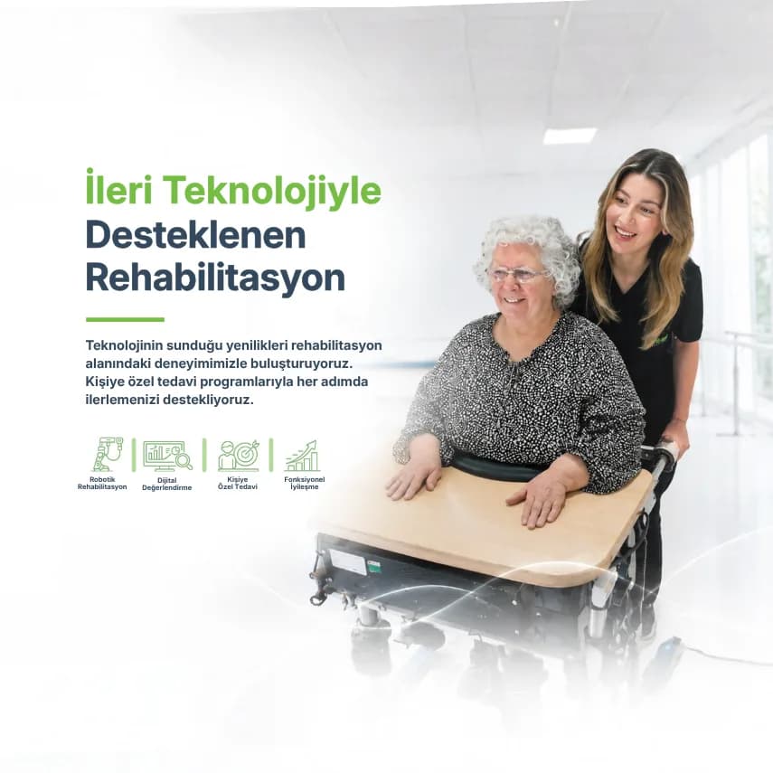
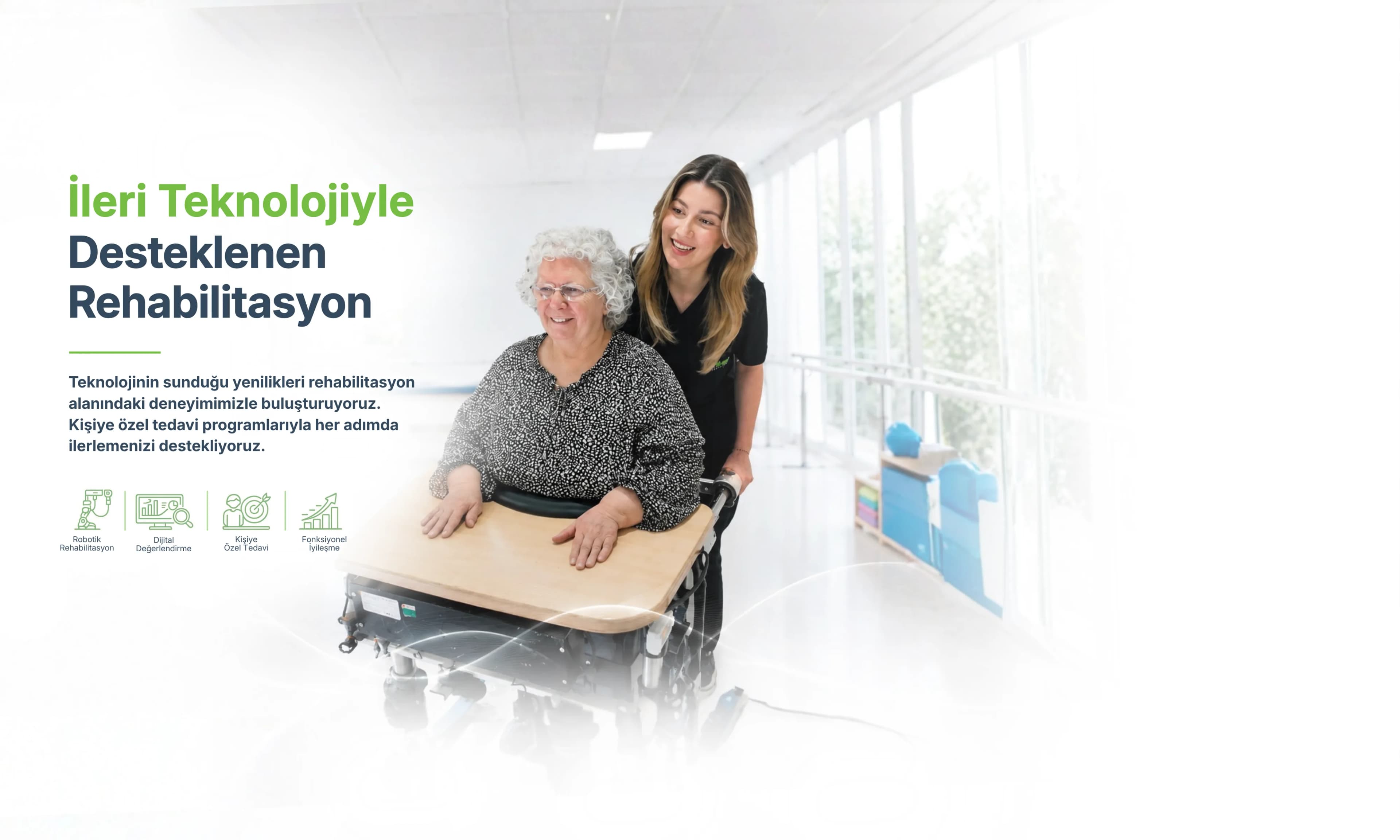
İnme, omurilik yaralanması, serebral palsi, multipl skleroz, Parkinson hastalığı ve ortopedik ameliyat sonrası dönemlerde robotik rehabilitasyon sistemlerimizden yararlanılmaktadır.
İlk değerlendirme seansı hakkında güncel bilgi almak için lütfen merkezimizi arayınız. Anlaşmalı sigorta kapsamındaki hastalar için sigorta koşulları geçerlidir.
Evet. Uluslararası hasta koordinatörlerimiz, yurt dışından gelen hastalarımıza konaklama, transfer ve tedavi planlaması konularında destek sağlamaktadır.